Manipulation of Photorespiration H-Protein Leads to Increased Biomass in Tobacco Plants

The effort to increase food production levels and, as a result, food security, has led us to pen a number of articles about the advantages the C4 photosynthesis over plants and crops that use the C3 photosynthetic system.

In short, plants that use C4 photosynthesis have the ability to concentrate carbon around RuBisCo, thereby reducing the competition between oxygen and carbon atoms for this important enzyme’s binding site. Reducing the amount of oxygen able to be bound by RuBisCo reduces photorespiration levels, photorespiration being an energy intensive process that converts oxygen into CO2.

Given the superior carbon-fixing efficiency of C4 plants and resultant superiority in growth speed and efficiency, efforts have been focused by a number of researchers and research organisations on transplanting C4 photosynthesis systems into important crops that rely on C3 photosynthesis for carbon fixation and growth.

However, a recent article published in the Plant Biotechnology Journal looked more closely at the photorespiratory process of the C3 tobacco crop and, particularly, whether the process can be made more efficient to the benefit of plant growth.

Introduction

The researchers first note that previous research which limited or removed the photorespiration process resulted in reduced plant growth and development, even though the aim of those experiments was to remove the inefficiencies in carbon gain caused by the process. They then write about research that assisted the photorespiratory process by increasing flow through the glycine cleavage system (GCS) which, promisingly, resulted in increased biomass in Arabidopsis thaliana.

The GCS is described as a set of three enzymes (P, T and L) plus a small lipoylated protein (the H-protein) which moves between and interacts with the P, T and L enzymes. Previous experiments have shown that over-expression of the H-protein can result in an increase in GCS activity and biomass accumulation amongst other positive effects.

Following on from the previous research, the researchers looked at the effects of different promoters in front of the H-protein gene to increase expressiond on the growth of tobacco plants, using the constitutive promoter CaMV 35S and the light regulated ST-LS1 promoter.

Results

The researchers took an A. thaliana H-protein cDNA and over-expressed the gene in tobacco plants behind either the CaMV 35S promoter or the ST-LS1 promoter. Lines from each construct were self-fertilised to create T2 lines which were then subjected to quantitative reverse transcriptase PCR (qRT-PCR) and immunoblotting to quantify gene expression, confirm H-protein overexpression and identify homozygous and null mutant lines. Further, normal levels of expression of the L, P and T genes were confirmed.

H-protein expression levels showed that expression behind the CaMV promoter resulted in three to seven times more H-protein than when expressed behind the ST-LS1 promoter.

The effect of over-expression levels of the H-protein on plant growth were then assessed. The plant lines were grown in a controlled environment for 26 days after which leaf area and chlorophyll fluorescence levels were measured. The researchers found that the CaMV lines showed a significant reduction in leaf area compared to control plants (up to 50% less), while those lines where the H-protein was expressed behind the ST-LS1 promoter had leaf area similar to or up to 26% greater than control. No significant difference between chlorophyll fluorescence levels was found.

H-protein overexpression fig 2b

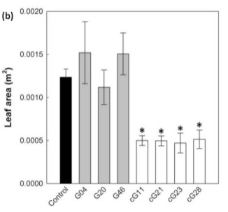
Figure 2b from article. Leaf area after 26 days of growth, demonstrating increase in leaf area in 2 of 3 ST-LS1 over-expression lines (in grey) and reduced leaf area in CaMV overexpression lines (white).

To analyse whether carbohydrate types and levels were a possible explanation behind the reduced leaf area, the researchers analysed carbohydrate profiles and found that whilst carbohydrate levels remained simlar between the CaMV lines and control, those transgenic lines had significantly lower levels of soluble sugars and increased starch accumulation. This alteration in profile was not seen in the ST-LS1 transgenic lines.

Lipoylation of the H-protein is essential to the function of the GCS, so the study looked at whether there was an alteration to lipoylation levels in the transgenic lines compared to control using immunoblotting of the lipoic acid groups in leaf and root tissue.What they found was that the lipoic acid levels in the transgenic plants were higher than in control. Whilst this makes sense given the higher H-protein levels in those lines, the greater levels of H-proteins in the CaMV lines compared to the ST-LS1 lines should result in a greater signal from the CaMV lines. However, the signal from both transgenic lines were similar, indicated that the extra H-proteins in the CaMV lines were not all active.

Given the negative effects of the CaMV promoter lines, the researchers chose to continue analysing the ST-LS1 plants further, testing the effects of the over-expression further under both glasshouse and field conditions.

Under glasshouse conditions, the transgenic lines showed increases in plant height, leaf number, leaf area and dry weight. Compared to control, above ground biomass of the transgenic lines were between 13% and 38% greater.

Glass house conditions are well and good, but the researchers wanted to see whether those increases were also possible under field conditions. So they performed two studies in consecutive years, the first being a small replicated control experiment using two transgenic lines which were grown for 25 days in a glasshouse before being moved into the field where they were assessed after 14 days. Once harvested, increases in biomass of both transgenic lines compared to control were again noted, resulting from increases in leaf number, height and total leaf area. Above ground biomass was 40% to 47% greater in transgenic lines compared to control.

The experiment was performed again the following year but this time using a larger, randomised block design. After being grown in the glasshouse for 33 days, the transgenic lines and controls were planted in the field and allowed to grow for a further 24 days and then harvested. Again, a significant increase in biomass was observed however one line, which had shown a significant increase over control in the previous year’s experiment, failed to outperform control. The other two transgenic ST-LS1 lines had increases in height (>18%), leaf area (>15%) and dry above ground biomass (>26%) compared to control.

H-protein overexpression fig 4

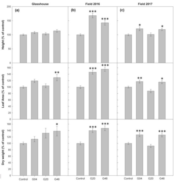
Figure 4 from article. Graphs depict differences in plant height, leaf area and dry weight between control and 3 ST-LS1 transgenic lines under greenhouse and two field experiements.

Testing whether increases in biomass were tied to changes in photosynthesis increases, the study analysed CO2 assimilation. They found that photosynthesis rates in ST-LS1 lines were increased compared to controls particularly under higher light intensities in glasshouse and field grown plants.

Finally, noting that conditions that trigger photorespiration are known to damage photosystem II, the researchers hypothesised that the increased photosynthesis rates in overexpressed H-protein lines may result from better protection of the photosystem from damage. To test the hypothesis, transgenic lines and controls were grown for nine days before being put in conditions of low O2 and continuous light – high photorespiratory conditions. They were subjected to these conditions for 24 hours before being returned to their original growth conditions and monitored for photosynthesis efficiency. The analysis found that photosynthesis efficiency was better preserved in transgenic lines compared to control.

Conclusion

The study finds that plants can have too much of a good thing when it comes to overexpression of the H-protein and GCS activity in tobacco plants. Whilst intermediate level of increased H-proteins resulting from gene promotion by the light-dependent ST-LS1 promoter resulted in increased biomass possibly due to improved photosynthesis rates and photosystem protection, too significant over-expression behind the constitutive CaMV failed for complete lipoylation of the extra H-protein, resulted in an altered carbohydrate profile and had reduced biomass compared to control plants. Importantly, growth under field conditions showed similar gains to those under controlled conditions.

The researchers have provided us with an alternative route to improving photosynthetic rates and crop growth rates than the more popular C4 engineering work. This study may provide either as an intermediate step towards greater crop production rates whilst C4 transformation research continues, be an improvement process that can employed in difficult-to-engineer plants or, should C4 transformation of C3 crops fail to become reality, improving photorespiration process could be one of many improvements in crop growth we see developed further in the future.

Researchers Discover How Rice Blast Fungus Spreads Inside Plant Tissue

Magnaporthe oryzae (M. oryzae), the causative fungus of rice blast disease, has the ability to (and does) destroy large swathes of cereal crops world-wide. Quite a long time ago we wrote about the possibility of using CRISPR to help control the disease by manipulating rice-plant genes to decrease susceptibility to the disease. The effort to increase resistance to rice blast was also mentioned in our post about the current status of genetic engineering’s promise to assist sustainable control of plant disease and our post on the possibility of increasing resistance through increased expression of the gene WRKY45.

Although progress towards increasing resistance to the disease is being made, better understanding the pathogen’s methods of infection and spread in turn informs methods of controlling and minimising the pathogen’s virulence.

The researchers behind a recent paper in Science delved into the methods of colonisation and spread used by the fungus on the background of previous research demonstrating that, once the fungus manages to invade the outer cuticle of the leaf, it suppresses plant immune responses. Concurrently, it produces hyphae that manage to spread to adjacent cells via a segment of constricted hyphae traversing plasmodesmata, rapidly infecting the tissue as result.

The Research Findings

The battery of tests performed by the researchers to better understand the fungus began with the use of transmission electron micrographs of rice sheath which confirmed that hyphae, normally measuring 5.0 µm in diameter, narrow to 0.6 µm in diameter to penetrate through the plasmodesmata, maintaining the integrity of the plasma membrane of the infected cells.

rice blast MAP kinase1

Figure 1A from article, showing the constriction of the invasive hyphae (IH) as it penetrates the rice cell wall (RCW).

Normally, callose is deposited to close plasmodesmata channels as an immune response in plants. This was visualised by the researchers but in cells that had been infected by the fungus, no callose was deposited 27 hours after infection. At 30 hours post infection, when initially invaded cells began dying, callose began to form at plasmodesmata but by this time the hyphae had entered adjacent cells. Callose then formed around the base of hyphae that had entered into the adjacent cell.

Testing the ability of the fungus to overcome the restriction on the size of molecules passing through plasmodesmata, mCherry expression vectors expressing either single or double-length mCherry proteins were inserted into infected and control rice tissue. In control tissue, single-length proteins were able to pass between cells while double-length proteins were excluded. However, in infected tissue, the double-length proteins were also able to pass between cells, indicating that the fungus is able to manipulate the size exclusion limit for long enough to assist hyphae spread. However, secreted effector proteins did not traverse the plasmodesmata into subsequently infected cells; effector proteins were limited to the initially infected host cell.

rice blast MAP kinase2

Figure 1G from article. Difference in movement of double mCherry proteins in plants infected and free from rice blast.

Investigating the control of hyphae growth, the researchers built upon earlier work that had shown that mutant variants of M. oryzae that knocked out a mitogen-activated protein kinase (Pmk1) failed to infect rice plants even when the plant leaves are wounded. To test this finding further, the authors of the paper created a Pmk1 allele that was sensitive to the analog 1NA-PP1, allowing the expression of Pmk1 to be manipulated in vivo. The study found that that when Pmk1 was turned off, the ability of the formed hyphae to constrict at the plasmodesmata entrance was reduced, preventing invasion of adjacent cells. What the researchers found was that in the presence of the analog the hyphae still formed terminal swellings at points where those hyphae met with the host cell walls but they were able to infect no further.

rice blast MAP kinase3

Part of figure 2A and figure 2C from article. Figure 2A demonstrates reduced ability for hyphae to invade adjacent cells when pmk1 expression is limited. Figure 2C highlights the swelling of hyphae when they meet the cell wall when pmk1 is inactive.

To confirm the role of the MAP kinase, a Green Flourescence Protein tag was added to the kinase protein to allow live imaging. Imaging showed that the gene was expressed at the time the fungus penetrates leaf cuticle and when the hyphae meet with plant cell walls just prior to those hyphae spreading to the adjacent cells.

RNA sequencing analysis was performed to compare gene expression levels when the Pmk1 gene was turned on and off. Inhibiting the gene resulted in the regulation of effector genes that are known to be involved in the suppression of plant immunity, as well as down-regulating a number of morphogenic regulators. To test whether it was the lack of host immunity suppression that was stopping hyphae from invading adjacent cells and not the Pmk1 kinase itself, the researchers suppressed host immunity simultaneously with Pmk1 suppression. What they found was even in the extreme test of killing plant tissue with ethanol and then rehydrating prior to inoculating the tissue with the fungus, suppression of Pmk1 expression still resulted in the hyphae being unable to infect adjacent cells, remaining trapped in the first host cell infected.

The result of these findings was the hypothesis that Pmk1 was critical to the fungus being able to constrict hyphae to cross plasmodesmata and invade neighbouring cells, the constriction being dependent on septin-dependent appressorium repolarisation, consistent with the effect Pmk1 gene expression had on the morphogenic regulators mentioned above.

To investigate the septin organisation effects of Pmk1 inhibition, a GFP tag was added to the Sep5 gene and imaged. What the researchers found was that the Sep5-GFP still accumulated where the hyphae met the cell wall but instead of forming septin collars at the site where the hyphae would cross the cell wall, the protein accumulated in a disorganised mass.

Conclusion

The results of these studies suggest that the Pmk1 MAP kinase pathway controls the ability of the fungus to use plasmodesmata as a means of moving hyphae from cell to cell, that ability relying on the constriction of the hyphae to a size that allows it to penetrate through these inter-cellular channels. Combined with the expression of effector molecules to suppress plant immunity that would otherwise see these channels blocked before the hyphae are able to invade, the pathway plays a significant role in the fungus’ virulence.

The conclusions drawn about the importance of the pathway in the fungus’ ability to infect and spread through rice plants must lead further research into methods of blocking the pathway in crops. Perhaps the use of RNAi targeted to this gene or host-derived endonucleases guided to the gene with CRISPR-based guide sequences could assist with application of this knowledge to a problem which regularly reduces the amount of rice produced in many parts of the world.

Great research!

Extending food storage at room temperature with ABA application

Food waste is a significant problem globally and one that we have written about previously in the context of wasted resources and climate change. Taking data from the Food and Agriculture Organisation of the United Nations, the authors of the paper we are looking at today report that the amount of food loss, being food successfully produced but which must be discarded before being sold to consumers, amounts to something in the vicinity of 45% of food produced globally.

A large portion of fresh food requires refrigeration in order to extend shelf life. However, refrigeration throughout the transportation and storage periods after harvest can be problematic to maintain. Further, cold storage post-harvest can effect the nutrient values of the produce and can effect the look, feel and taste at the time of purchase and consumption.

The researchers behind a paper published recently in the Plant Biotechnology Journal studied the physiological and hormone profiles of detached cabbage leaves stored at room temperature and in cold conditions in an attempt to characterise differences between the two types of storage conditions and identify those differences that could be the cause of the increased senescence of those stored at room temperature.

Given the significant amount of research that has characterised abscisic acid (ABA) as a plant hormone which affects responses to stress (we’ve previously reported on a mini-review of the role of ABA in plant stress response here), the researchers further studied the response of the detached cabbage leaves when ABA or pyrabactin was applied to the leaves and then stored at room temperature. Pyrabactin is a synthetic sulfonamide which can mimick the effects of ABA but is less photosensitive, possibly more selective in its effects on plants and probably easier to apply by farmers.

The effects of cold storage compared to room temperature storage

The paper details that cabbage leaves were detached and placed either in storage at 4°C (cold storage) or at 25°C (room temperature) and monitored over 10 days.

After 10 days in storage, leaves stored at room temperature lost half their initial weight compared to only 20% when kept in cold storage.

All other methods of analysis were performed on the separated apical, medial and basal strips of leaves stored at either temperature.

Water loss from apical strips of cold stored leaves was less than those stored at room temperature, while there was little difference in water loss between the basal strips.

Photosynthesis efficiency was significantly reduced across all strips stored at room temperature after 4 days of storage compared to no change under cold storage. The medial and basal strips had decreased chlorophyll and carotenoid levels under room temperatures, characterised by yellowing of the leaves and water loss.

Leafy Vegetable Figure 1

Figure 1 from article. Differences in relative water content (RWC) and chlorophyll levels in apical, medial and basal strips of cabbage leaves stored at room temperature and 4°C.

Hormonal profiling showed significant changes between the two treatments, with decreases in levels of a number of cytokinins in leaves stored at room temperature and ABA contents increasing to a greater extent in leaves stored at room temperature compared to those stored in the cold.

The effects of applying ABA and/or Pyrabactin on senescence

Given the difference in ABA levels in leaves stored at room temperature, ABA and/or Pyrabactin (Pyr) was applied to  leaves to test whether it had any effect on the deterioration of leaves stored at room temperature. Either ABA alone, Pyr alone or a combination of ABA and Pyr were applied to leaves every second day for eight days and then analysed in comparison to control leaves.

Membrane stability, analysed via a number of different methods, demonstrated that each of the three treatments increased membrane stability compared to control. Despite increased stability, photosynthesis efficiency was not significantly different between experiment and control.

Leafy vegetable senescence

Figure 3 from article. Water retention, chlorophyll, malondialdehyde and Membrane Stability Index measurements compared between leaves at the day of harvest and between each treatment after 8 days.

Hormone profiling did however show some significant changes caused by the ABA/Pyr application. Endogenous ABA levels were doubled in treated leaves with the application of ABA (but not Pyr alone) and several cytokinins showed increased levels with treatment.

Transcriptomic analysis was then performed on the apical strips of control leaves and each of the three treatments. Despite the doubling of endogenous ABA after treatment, ABA transcription showed no marked changes but ABA catabolism and ABA release from reversible conjugation were down-regulated. ABA transporters in the cytosolic membrane were expressed while those in the vacuole were not expressed, likely due to the increase in ABA from the application and therefore reducing sensitivity to the hormone.

Ethylene metabolism and regulation showed some significant changes with the increase in ABA content. At the end of the treatment period the presence of the precursor to ethylene, AAC, was halved in treatments containing ABA with genes encoding the syntheses of ACC and ethylene both down-regulated. Negative regulators of the ethylene response were also down-regulated. Pyr treatments showed little or no effect.

The manipulation of hormones by the ABA/Pyr treatments suggested to the researchers that the treatment may delay senescence in the cabbage leaves. Many photosynthesis genes were up-regulated as a result of treatment, as were genes coding calvin cycle enzymes. Genes involved in the reduction of reactive oxygen species were also up-regulated, while genes involved in maintenance of cell walls and membranes were also found to be up-regulated by the ABA/Pyr treatment. These findings gave plausibility to the possibility that senescence was being delayed in the treatment group.

In a possible trade off for the delay in senescence, the ABA/Pyr treatments appeared to down-regulate the defence mechanisms of the leaves to biotic stress. Treatment with ABA resulted in reductions in salicylic acid-related defence responses.

Conclusion

The researchers state in the conclusion that ABA’s role in regulating senescence isn’t overly surprising given its role in assisting plant responses to abiotic stress such as drought, a stress imposed on leaves when they are detached from the plant. Previous research has indicated that the role ABA plays in the stress response of leaves depends on the age of the leaf; ABA stimulates senescence in older leaves while resisting senescence in younger leaves. By applying ABA to leaves immediately post-harvest, the treatment promoted homeostasis and assisted the leaves to resist the senescence observed in the untreated leaves.

The findings of the paper suggest a plausible method of reducing food loss post-harvest. However, the practicability of applying ABA to leaves immediately post-harvest remains to be seen and the transferability of the findings to other fresh produce will be interesting to see. Also of interest would be the difference in senescence delay between ABA application and cold storage to determine whether there is any ability to reduce reliance on cold storage.

The research should lead to further investigations into the role of the individual components of the senescence response of harvested plants and how significant a role manipulating ABA responses can play in securing food availability. Further, the applicability of transgenic methods in generating the same responses without the need for exogenous application of ABA could be a research target which could generate interesting results.

CRISPR-Derived Plant Resistance to RNA Viruses

The fight against crop damage from biotic and abiotic stresses has led and continues to lead research in the fields of soil science, stress signaling and hormone responses, plant immunology and immunology more generally.

One breakthrough application from the discovery of RNA interference (RNAi) is the ability to use this method of host protection to specifically target particular pathogens that a plant may be susceptible to. The most prominent example of the application of this technology was its use to save the papaya industry from the Papaya Ringspot Virus (a great explanation of the virus and the use of transgenic technology to control it can be found here).

The discovery and development of CRISPR technology has also been used to tackle plant stress issues in multiple ways, whether that to be to direct endonucleases to invading genetic material or as a tool to insert sequences that allow the plant to generate its own resistance abilities.

However, as the researchers behind a recent paper in the Plant Biotechnology Journal state, whilst the ability to protect plants against DNA-based viruses using CRISPR is increasing, the tool hasn’t found implementation in the effort to control RNA viruses primarily due to it being limited to the restriction of target DNA.

Therefore, the researchers took some recent findings into the ability of some variants of the Cas9 endonuclease (the common nuclease used with CRISPR) which had the ability target RNA sequences within prokaryotes to test whether they could be used to develop resistance to RNA viruses in plants.

The Study

Using cucumber mosaic virus (CMV) and tobacco mosaic virus (TMV) as the two RNA viruses to be targeted, the researchers took the Cas9 protein from Francisella novicida (FnCas9) and used it in combination with guide RNAs directed toward a variety of targets within the CMV and TMV genomes, then assessed whether the construct had any effect of infection rates and severity compared to control plants.

Controlling CMV

The first part of the experiment focused on CMV, a virus with a genome of three parts, each with similar sequences. The FnCas9 was to be directed to 23 sites within the genome selected by the researchers and complementary sequences to those sites were individually synthesised and inserted into separate vectors also containing the sequence for the FnCas9 nuclease.

Constructed vectors were inserted into Agrobacterium which was then injected into a bottom quarter of 25 day old N. benthamiana leaves. A control vector was injected into the opposite bottom quarter of the same leaf and the CMV infection was inoculated in the top half of the leaf.

CRISPR RNA Virus1

Figure 1(c) from article, demonstrating the inoculation sites of the experimental vector, control vector and CMV on subject leaves.

Five days later, discs were cut from the bottom quarters of the leaf for analysis of infection rates. The analysis was performed by quantifying virus RNA presence using Reverse Transcription Quantitative Polymerase Chain Reaction (RT-qPCR) and found that the quantity of CMV RNA in the discs containing the FnCas9 + guide RNA was between 40% and 60% less than that contained in the control discs.

Within the same experiment the researchers toyed with different PAM spacers but found that there didn’t seem to be any variability between the different spacers used and the performance of the FnCas9 construct.

The three RNA target sequences which resulted in the best virus resistance were selected for further testing. FnCas9 constructs containing the individual RNA targets and controls were again transiently expressed in N. benthamiana plants and the following day the plants were challenged with the CMV virus. Two weeks later, control plants showed symptoms of CMV infection while severe infection symptoms were avoided in the plants inoculated with one of the three FnCas9 vectors. ELISA-based quantification and RT-qPCR quantification of viral RNA again showed reductions of between 40% and 50% compared to control plants.

CRISPR RNA Virus2

Figure 2 from article showing the difference in viral effects between control and transgenic plants (a) and comparison in viral load using ELISA (b) and RT-qPCR (c).

Controlling TMV

The researchers took a different tact to researching the ability of the FnCas9 constructs to control TMV. They attached a Green Fluorescence Protein to the virus and then targeted the virus with FnCas9 constructs directed to three selected sequences within the TMV genome. Accordingly, expression of TMV would result in detectable fluorescence, whilst the suppression of the virus would result in a reduced amount of fluorescence.

One week after inoculation with the virus, bright green fluorescence was observed throughout the control plants but treated plants showed significantly reduced fluorescence comparatively. Again, RT-qPCR confirmed the reduced virus load in the treated leaves.

Researching the Mechanism

The results appear to show that the FnCas9 plus RNA guides are working well in repressing the RNA viruses. To check whether the synthetic constructs are the true cause of the effect seen and, if so, to delve into how the construct is working, the researchers performed a number of tests.

The first test used immunoprecipitation targeted at a flag placed at the N-terminus of the FnCas9 protein to capture and purify the FnCas9 + RNA guide. When the purified proteins were sequenced and amplified using RT-PCR the CMV virus was also detected, indicating the guide RNA was associating the viral RNA and therefore the FnCas9 protein was being successfully directed to the viral RNA genome.

To check whether the FnCas9 was necessary to the inhibition of the viruse, the FnCas9 was replaced with a GUS reporter gene. When expressed with the guide RNA the CMV virus progression was no longer repressed by the synthetic construct. Therefore, the FnCas9 protein was required for virus repression.

The next method used to check the mechanism was to add a nuclear localisation signal to the FnCas9. Restricting the protein to activity only within the nucleus of the plant cells again resulted in reduced virus repression compared to that seen previously.

Point mutations were then made to the active regions of the FnCas9 to disrupt its endonucleolytic activity.  Interestingly, the disruption to the ability of the protein to cut the RNA sequences made no difference to the ability of the construct to reduce viral load and disease severity.

The last test the constructs were put to was test whether the ability to bind the viral RNA effected the productivity of the construct. Mutations were again made to the construct, this time at the RNA-binding arginine-rich motif (ARM) of FnCas9. The inability of the FnCas9 protein to bind to the RNA resulted in reduced ability of the construct to repress the RNA virus.

Accordingly, the ability of the FnCas9 synthetic constructed directed to the two RNA viruses to curtail infection relied not the endonuclease cutting the viral RNA but actually relied on its ability to bind to the viral genome, an unexpected result which the researchers later gave the name CRISPR Interference (CRISPRi) in the discussion section of the paper.

Stability of Resistance

Transient expression of the FnCas9 constructs resulted in repression of virus load, but to be of assistance to crop protection, stable repression of virus is required.

Therefore, the three best performing constructs targeting CMV were used to transform Arabidopsis thaliana plants. Homozygous T2 plants for each construct were created and selected for challenge with CMV infection. While control plants showed severe effects of infection two weeks after being infected, transgenic plants mild symptoms in most lines with two lines having no obvious symptoms. ELISA-based quantification confirmed the virus inhibition.

The same lines were continuously harvested until the sixth generation was produced and then challenged with infection.  The randomly selected lines showed a stable resistance to the virus .

Conclusion

The study demonstrates a novel method of reducing the effects of a couple of important RNA viruses that can have dramatic impact on plant viability and productivity. Whilst the logic behind the study is sound and seemed destined to work given FnCas9 had previously been demonstrated to target RNA, the mechanism by which the endonuclease repressed was not anticipated.

Acting effectively like RNAi, the FnCas9 seems to block the translation of the virus’ RNA genome, halting its ability to infect the plant cells. Further, constitutive expression was shown to be stably inherited in subsequent generations.

The researchers point out in the discussion section of the paper that this strange method of reducing viral loads could have some advantages, such as a reduction in the production of mutant forms of the virus from cleaved RNA strands escaping the cells. However, it does have a similar limitation to RNAi in the creation of evolutionary pressure on the virus to mutate to avoid the repression of the site of the genome targeted by the FnCas9.

Although the same problem of the “arms race” between virus and plant arises in the context of this research, the findings certainly give some hope that CRISPR can be used to reduce the effects of viral damage to crops.

Assembling plant RuBisCo in E. coli

Optimising photosynthesis is a significant goal for those researching ways to secure the human food supply on a planet that will continue to see the emergence of threats to agriculture from climate change. We’ve written a number of articles in this vein, predominantly on the effort to be able to engineer C4 photosynthesis into major crops that use the less efficient C3 photosynthesis (see here, here, here and here for a few examples).

In the research article “Plant RuBisCo assmbly in E. coli with five chloroplast chaperones including BSD2″ published in Science, researchers from the Max Planck Institute of Biochemistry sought to install a functional RuBisCo enzyme from our favourite model organism, Arabidopsis thaliana, into E. coli, the model bacteria with a short generation time.

The researchers explain that the purpose of attempting to install plant RuBisCo into the bacteria is to assist the effort seeking to improve the catalytic efficiency of the enzyme, allowing it to be studied closely in conjunction with genetic engineering to hasten the results of this line of research.

The genes that transcribe the subunits of RuBisCo have been known for some time and the arrangement of eight large subunits (RbcL) and eight small subunits (RbcS) is well characterised. But as with the ongoing quest to understand protein folding patterns, the process by which the holoenzyme comes to be in its final conformation, which supplementary proteins are required to assist the assembly of the enzyme and therefore which supplementary genes need to be expressed for the functional formation of the enzyme, are not well understood.

By attempting to build the enzyme from scratch, we learn more about the necessary genetic components required to build it, as these researchers found.

The Study

The researchers first attempted to express A. thaliana RuBisCo in E. coli by transferring the RbcL and RbcS genes along with two additional factors, RbcX and Raf1 (the functions of which were further elucidated later in the paper), but this attempt was unsuccessful.

Therefore, they attempted the feat again using three separate plasmids, one containing the RbcL and RcbS components, another containing chloroplast chaperonin proteins (C60αβ/C20), and a final plasmid containing four assembly chaperones (Raf1, Raf2, RbcX and BSD2). Using separate plasmids allowed the researchers to test which components were required to form functional enzymes. Expressing all genes resulted in the formation of proteins that migrated on polyacrylamide gel electrophoresis at the same position as AtRuBisCo and which had a similar affinity and carboxylation rate of CO2 as native RuBisCo. Plasmids that did not express either sets of chaperones failed to form proteins that migrated to the same position.

Accordingly, the RbcL and RbcS genes plus the genes encoding the various chaperones transferred into the E. coli appears to have resulted in the expression of functional RuBisCo.

Chaperone Requirements

Being able to functionally express RuBisCo from a higher order plant in E. coli is a significant step forward, but understanding the purpose and need for each of the chaperones remains outstanding. To probe a bit deeper into the requirements for functional RuBisCo enzymes, the researchers varied and substituted a number of the genes and monitored the outcome.

The first reported variation was to substitute the C60αβ/C20 chaperones with GroEL/GroES, endogenous chaperones from E. coli. The researchers found that even overexpressing the endogenous chaperones couldn’t match the level of functional expression of RuBisCo compared to when A. thaliana chaperones were used. Cpn60α and Cpn60β were both required for efficient expression of the enzyme, while expression of a plasmid without C20 still resulted in the formation of RuBisCo, albeit with a lower rate of expression. Overexpressing GroES in the absence of C20 resulted in the production of RuBisCo with similar efficiency as when C20 is coexpressed.

RuBisCo in E Coli1

Figure 2 from article. Chloroplast charperonin requirements to form functional RuBisCo. Only when the RbcL, RbcS and folding chaperones were expressed with C60αβ/C20 (lane 2) or with C60αβ/GroES (lane 9) did RuBisCo successfully form.

In relation to the Raf1, Raf2, RbcX and BSD2 factors, the researchers deleted each of these in turn to test the effect on the final outcome of the enzyme to gain further insight into the function of each.

Deleting Raf1, Raf2 or BSD2 resulted in no RuBisCo production. Previous research has demonstrated that Raf1 works to assemble the RbcL subunits into RbcL8, whilst the function of Raf2 remains to be diagnosed.

RbcX deletion didn’t completely halt RuBisCo production but reduced assembly rates by 50% to 60%.

BSD2, what does it do?

The research delved much deeper into the function of BSD2, a gene that isn’t contained in all photosynthetic organisms and appears to have evolved subsequent to the endosymbiosis of the organism that led to chloroplasts, unlike the other three chaperones.

It was theorised in the paper that, given the possible evolutionary origins of the protein, that it may be related to the importation of the RbcS subunits into the chloroplast. When RbcS wasn’t expressed, a RbcL8/BSD2 complex formed. When RbcS was reintroduced, the RbcL8/BSD2 complex no longer formed; instead the RuBisCo RbcL8S8 enzyme formed. Omission of RbcS and Raf1 resulted in the lack of formation of the RbcL8/BSD2 protein, suggesting that Raf1 plays a role in the formation of this intermediary phase of the holoenzyme.

Given that it appears BSD2 plays a role late in the construction of the RbcL8S8 enzyme, the researchers tested whether altering the BSD2 and RbclS expression would have similar affects on the cyanobacterium Synechococcus elongatus. Without the expression of SeRbcS, SeRbcL and BSD2 formed a SeRbcL/BSD2 complex which, when SeRbcS was reinstroduced, resulted in fully formed SeRuBisCo, similar to what happened with the Arabidopsis based genes when expressed in E. coli. Therefore, this experiment helps to confirm the hypothses that BSD2 play a late but significant role in the final formation of RuBisCo.

RuBisCo in E Coli2

Figure 6D from article. Model of protein folding sequence and required chaperone involvement to form functional RuBisCo.

Taking our knowledge of this process a step further, the structure of the BSD2 protein and how it may interact with RbcL to form a RbcL8/BSD2 complex were analysed.

BSD2 imaging showed that it forms a hairpin structure that centres around two zinc atoms. Imaged as part of the RbcL8/BSD2 complex, the crystallised structure shows that two BSD2 molecules are bound within antiparallel RbcL2 units, appearing to join the two RbcL subunits together. One of the zinc-containing regions of the BSD2 interacts with the C-terminal end of one RbcL unit while the other zinc-containing region interacts with the N-terminal of the adjacent RbcL subunit. It appears that the conformation of the RbcL subunits when interacting with BSD2 allows RbcS subunits to access and bind to the required sites on RbcL. Mutating the BSD2 at contact sites resulted in a loss of function of the protein.

Conclusion

This piece of research will hopefully assist to increase efficiency in RuBisCo research, research which aims to increase photosynthesis efficiency with resultant improvements in crop yields and food security. The research is somewhat Feynmann-esque, referring to the note scribbled on his chalkboard when he passed that:

“What I cannot create, I do not understand”. Richard Feynmann, February 1988

Although this research is useful in its application for further research, the attempt to recreate the enzyme from the scratch also helps us to further understand the function of those genes and moving us closer to the depth of knowledge Feynmann aspired to.

Great research resulting in a method that we expect to see in use and built upon in future.

Host-delivered RNAi to control Wheat Leaf Rust

Wheat is a major staple food worldwide. Accordingly, threats to wheat production pose a significant risk to global food production and security. One major threat to wheat production is Wheat Leaf Rust (“WLR”), a disease caused by the fungus Puccinia triticina which has the ability to cause severe yield losses. Of further concern, the application of fungicides to control the disease can lead to the development of reduced sensitivity or resistance to the fungicide, let alone the economic and environmental impacts that fungicide use entails.

Given the serious threat that WLR entails and the problems with using fungacides, genetic studies have been used to identify a number of resistance genes which have been used to breed cultivars with greater resistance to the fungus. However, the use of resistant cultivars in monoculture applies significant pressure on the fungus to evolve and develop new methods of virulence.

The authors of the paper being presented today outline these important problems noting that, when combined with the effects of a changing climate, disease outbreaks are at risk of increasing and further threatening food security.

A Potential Solution?

After discussing the life cycle and infectivity of the fungus, the researchers behind the paper published in the Plant Biotechnology Journal theorised that by using the increasing genetic knowledge about the pathogen, particularly the genes implicated in their virulence factors that enable it to infect wheat plants, it may be possible to target and repress those genes using RNA-interference (RNAi).

We have written about RNAi previously in the context of RNAi sprays as a possible alternative to traditional pesticides, potentially being used in Bioclay-based pesticide and in transgenic plant pathogen resistance.

The researchers outline the possibility of using host-delivered RNAi (“HD-RNAi”), inserting genetic material into the plant that will translate into a double-stranded piece of RNA complementary to one of the virulence genes of the pathogen. When the dsRNA piece is cleaved into small interfering RNA (siRNA) duplexes in the plant and delivered to the pathogen, the siRNAs bind to the transcripts of the complementary virulence gene in the parasite and interfere with their further translation.

Using recent studies that characterised virulence genes of rust fungi, the researchers chose to target the Pt Map-kinase (PtMAPK1) and Pt cyclophilin (PtCYC1) genes, attempting to silence their translation and discover whether effective silencing of these genes will afford any protection to the plants against the fungus.

Results

HD-RNAi constructs targeting each gene were transformed (separately) into immature embryos of a WLR susceptible wheat cultivar via projectile bombardment, creating 14 transgenic To lines harbouring the hp-PtMAPK1RNAi construct and 20 To lines aimed at silencing the PtCYC1 gene. These lines were selfed to produce T1 seeds. Polymerase chain reaction (PCR) was used to verify successful insertion of the constructs and reverse-transcription PCR (RT-PCR) was used to verify transcription of the construct. Furthermore, there was no effect on phenotype seen in the transformed lines.

The transformed lines and a set of controls were then challenged with the Pt urediniospores when they were two weeks old. 12 days after being challenged, the plants were scored on a 0 to 4 scale depending on the size and abundance of uredinia, with 0 being little to no infection and 4 being highly susceptible.  A total of 13 of the transformed lines (6 of the PtMAPK1, 7 of the PtCYC1) showed varying resistance to the fungus compared with the controls. Those lines with the greatest levels of resistance showed symptoms of infection between 1 and 3 days later than the control lines did.

LWR Fig 1

Figure 1 from article showing WLR infection levels in select transgenic lines compared to control (on the left).

To further investigate whether the HD-RNAi were the cause of the difference in susceptibility to the fungus, the researchers looked at the rate of fungal growth within the leaves of the plants by comparing the ratio of fungal DNA to plant DNA between the transgenic lines and the control lines. Analysis showed that the amount of fungal biomass was reduced by nearly 80% in each line compared to the controls. Accordingly, it appears that the HD-RNAi constructs have managed to interfere with the fungus’ ability infect and grow in the plant.

Using the quantification of fungal biomass to group the plants with differing levels of WLR resistance, the connection between the level of resistance and the severity of disease was tested. It was found that there was a close relationship between the two criteria, with the lowest disease severity correlating with the lowest ratio of fungal biomass to plant biomass.

LWR Fig 5

Figure 5 from article. (a) Comparison of fungal and plant biomass within infect control and transgenic wheat lines. (b) Transcript levels of genes targeted by the RNAi construction compared to control. (c) Confocal microscopy of infected cells.

To test whether the HD-RNAi constructs were actually interfering with the targeted fungus genes, quantification of transcript levels of the fungus genes was performed 5 days post infection of both transgenic and control lines. Transcript levels of both target genes was reduced by between 41% and 65% in transgenic lines compared to controls, and the reduction of transcript levels of each particular gene was specific to the transgenic line expressing the HD-RNAi construct targeting that specific gene. Further, the measured transcript levels correlated with the level of disease severity in the plant, demonstrating a link between the reduction in target transcript, level of fungal biomass and disease severity.

Finally, three transgenic lines from each HD-RNAi transformation showing increased disease resistance were selfed to obtain T2 generations of each. PCR and RT-PCR confirmed the presence and translation of the gene construct in that generation and the plants were challenged with Pt urediniospores when they were two weeks old. Similar to the T1 generation, infection scores were lower in the transgenic lines compared to control lines, with a similar delay in onset of symptoms previous described.

The same analysis of fungal biomass and transcript abundance was measured, again with similar findings compared to the T1 generation.

Discussion

The results of the research are promising, and the discussion section of the paper explored the possibility of targeting more than one virulence gene in any one transformation event or targeting different virulence genes as we learn more about the genetics of the fungus.

Further discussion centres on how the HD-RNAi constructs are delivered to the pathogen, a process not fully understood. It is hypothesised that the HD-RNAi transcripts may enter the pathogen via vesicle-mediated transport across the haustorial interface. However, the delivery method requires further research to confirm this and to confirm how inter-kingdom RNA is able to interfere with one another.

Why there was a variation in resistance levels amongst the different transgenic lines was also discussed, with hypothesis such as incomplete knockdown of the target gene, variability of gene expression levels and target gene transcript turnover rates.

Conclusion

The study builds upon our growing knowledge of plant and pathogen genetics, pairing the two with our understanding of RNAi to explore the possibility of controlling a significant and increasing threat to wheat production and avoiding the fiscal and environmental problems that come with increasing fungicide use. The results of the study are promising and should direct further research. This further research may consider whether different pathogen genes can be targeted, whether targeting different combinations of genes will have greater effect, and testing whether the problem of forced evolution of the pathogen to overcome our advances will mean that our crop defence strategies will still require us to employ agricultural practices to reduce the evolutionary pressure our genetic strategies apply.

A great study with possible applicability to a range of pests in other important crops.

CRISPR-S: Quick and Cheap Selection of Transgenic Rice Plants

The CRISPR/Cas9 (Clustered Regularly Interspaced Short Palindromic Repeats) genome-editing technology is, by itself, a significant recent addition to the biologists’ toolkit. The tool, which gives researchers the ability to direct gene edits (whether that be single point mutations, deletions of a chunk of the genome or the addition of new genetic codes) to specific sections of the genome, has seen tremendous uptake in use both in systems biology research and genetic manipulation studies.

Not only does the tool have the impact of a sledgehammer with the precision of a laser, but manipulations of the tool have resulted in a swathe of CRISPR-derived tools that either improve the tool in some specific way or alter the effect that the nuclease has on the genome being edited.

A letter published in Wiley’s Plant Biotechnology Journal in August this year described a modification of the CRIPSR tool to allow easy identification of successfully edited rice plants and simple identification of second generation plants which, while having the desired genetic modification, no longer contained the piece of transfered CRISPR/Cas9 genetic material within the genome.

The CRISPR Modification

The paper starts with the researchers noting that in the course of editing genomes in plants there are two steps that need to be successfully completed.

To edit a plant genome, the CRISPR/Cas9 genetic material needs to be transferred and inserted into the genome to be edited. This is usually performed with Agrobacterium tumefaciens which randomly inserts the CRISPR/Cas9 sequences, promoter etc into the plant genome. When the sequence is subsequently expressed, the guide RNA and the Cas9 nuclease are expressed which in turn go on to attach and edit the desired part of the genome.

For this first step to be successful, the researchers not only need to ensure that the required modification is going to be made, but must also identify the regenerated plants that have been successfully modified. Although the rate of successful editing can be quite high it is not usually 100% effective and the process of confirming a successful edit within the first generation of the modified plant can be time consuming and expensive in itself.

Once the successfully modified plants have been identified, the CRISPR/Cas9 gene insert must be removed from subsequent generations of the plant so that only the modification to the gene of interest remains. Again, this can require time consuming and expensive genetic screening processes to identify the successfully transformed, but CRISPR/Cas9 gene-free, plants.

CRISPR-S

The researchers who penned the paper theorised that it may be possible to create a gene insert that self-reported whether it was successfully introduced by causing a change in phenotype (aside from the change being introduced by the CRISPR element itself). To do this they proposed that, by introducing a gene segment into the plant that contained the CRISPR/Cas9 sequence plus a sequence that transcribed a piece of hairpin RNA complementary to some other gene of known function, the result would be the required genetic modification plus RNA interference of a marker gene that could be used for identification of successful transformation.

CRISPR-S 1

Figure 1 (e) from the article demonstrating the successful transformation of the gene in bentazon-susceptible plants (top left), the transgene-silenced and therefore resistant plants (top right), the second generation plant with the edited cadmium transport gene but lacking the CRISPR transgene, therefore regaining bentazon resistance (bottom left), and the edited second generation plant with the transgene remaining in situ (bottom right).

To demonstrate the theory they sequenced a piece of DNA to insert into rice plants. The DNA being inserted contained a CRISPR/Cas9 segment which directed an edit to a gene which encoded a cadmium transporter, plus a segment of DNA encoding a piece of hairpin RNA that, if successfully inserted into the genome of the plant, would result in the plant being susceptible to the herbicide bentazon. Bentazon susceptibility would then be the marker for successful transformation of the first generation of plants (which would assume successful editing of the gene for cadmium transportation) and also serve as a marker for those second generation plants that had lost the CRISPR gene segment given that the lack of presence of that gene would then restore the plant’s resistance to bentazon.

Testing the theory

The researchers created the sequence and inserted it into japonica rice genotype Xidao #1 via Agrobacterium tumefaciens. The tillers of the first generation of plants were divided and grown as two subplants, one of which was sprayed with bentazon, the other which wasn’t (meaning that if one subplant was susceptible to the herbicide and died, thus confirming successful transmission, the other subplant also with the successful transformation remained). Of the 96 independent transgenic events, 29 subplants were susceptible to the herbicide with 67 subplants unaffected.

CRISPR-S 3

Figure 1 (b) from the article demonstrating the bentazon resistant (and therefore unedited) first generation plant (left) and susceptible (and therefore edited) first generation plant (right).

The 29 subplants with the RNAi affecting bentazon resistance were then sequenced at the site of the cadmium transporter gene to test whether the gene segment had not only created the hairpin RNA but had also edited this particular gene. The researchers found that all of the plants displaying the susceptibility marker also contained the desired gene edits at the cadmium transporter gene.

Thirdly, the researchers used quantitative real-time PCR to test the abundance of the Cas9 and the bentazon susceptibility genes to check whether it was their gene construct that was causing the changes being noted. They found that in the susceptible offspring the transcripts of the Cas9 gene was significantly higher and transcripts of the gene that normally confers resistance to bentazon was significantly lower (causing the susceptibility to the herbicide) than those found in the resistant offspring. Thus, it would seem that the gene construct had successfully resulted in transcription of the Cas9 nuclease and the RNAi construct, the latter reducing the transcript abundance of the bentazon resistance gene.

To test whether their marker construct could also be used to distinguish transgene-free second generation rice plants they created 16 lines (72 seedling each) from the successfully edited first generation plants. They again sprayed the plants with bentazon with the hypothesis that those generations which had lost the transfer DNA containing the CRISPR/Cas9 and RNAi gene segment would regain their resistance to the herbicide. Of the plants that died, subsequent analysis proved that the transgene DNA remained in the genome, while the resistant plants had lost the transgene segment in the recombination of genes during the creation of the second generation.

Conclusion

It appears that these researchers have created a method, combining CRISPR/Cas9 and RNAi, to enable easy identification of successfully transformed plants and to then confirm that the transgene component has been removed in subsequent generations. This is particularly useful in those countries where current legislation does not consider plants that have been edited to remove a section of DNA (as opposed to being edited to add a piece of DNA) as “genetically modified”, therefore avoiding regulatory control. Being able to easily confirm that gene deletion has occurred and then confirm that the transgene has been removed will reduce the time and cost associated with confirming the modifications using gene sequencing.

It will be interesting to see if the process can be used in other plants. The time and expense spared could see the speed of research projects increase and open up the technology for projects that lack funding to allow for repeated primer creation and PCR-based sequencing to confirm genetic alterations.

A Study on Studies that Report Adverse Effects of Transgenic Food & Feed

Our policy at the Legume Laboratory is to report on the latest science surrounding the way we feed ourselves and possible methods of feeding a growing population in a world of limited resources and escalating environmental pressures.

Although we have a keen interest in the public discourse surrounding genetically modified crops, delving into popular arguments against their use falls foul of the line we have drawn in the sand for what we will report on.

However, an article in the Plant Biotechnology Journal reviewed a number of studies that are usually cited by proponents of the anti-GMO movement as evidence of the adverse health effects of these crops. Although this is usually one tenet of a larger argument against the use of genetically modified food, the use of published scientific papers to support part of the argument opens it up the rigors of scientific critique. This review gave us the ability to write about the subject.

Which papers were reviewed?

Before deciding which papers were to be reviewed, the authors of the review point out the noticeable foible whenever reading a commentary on genetically modified crops – that the only commonality between the variety of “GMO” foods is the process of creating them. Although transgenic crops may be created by the same process, the type of modification made or the trait inserted into the crop are individually known as ‘events’. The paper reports that while the FDA has reviewed 153 individual events, the majority of studies that report negative effects and are the focus of negative commentary relate to relatively few events. Although the studies reporting adverse effects are highly publicised, studies reporting no safety concerns garner little public attention.

In selecting articles for review, the researchers took the citations listed in four review papers which concluded that there are negative effects related to transgenic agriculture (Domingo and Bordonaba (2011); Dona and Arvanitoyannis (2009); Magana-Gomez and de la Barca (2009) and Seralini (2011)) plus those cited by the organisations ‘GM Free USA’, ‘Coalition for a GM Free India’ and ‘GM Watch’. Of these, studies not published in scientific journals or which didn’t evaluate whole food/crops but only pure proteins were excluded.

The result was 35 studies which met the criteria. The authors make a number of observations about the selected studies, including that:

  • they make up fewer than 5% of the total number of studies assessing the safety of transgenic crops;
  • nearly half (43%) of the studies looked at a herbicide resistant soy bean the product of one particular transgenic event;
  • 23% of the studies focused on another single event, an insect-resistant maize;
  • 9% studied crops not commercially available;
  • 20% of the studies did not indicate which transgenic event was being evaluated;
  • 31% of papers were authored by the Malatesta group at two particular Italian universities;
  • Only one article was published in a high ranking journal – (Ewen & Pusztai (1999)). The article was published with an analysis written by the editors which elucidated several significant criticisms of the study. The editors also commented that by publishing the article they did endorse the study or its conclusion but instead thought that there was a public interest in publishing it so that it could be evaluated by other scientists and to avoid cries of suppression from scientists claiming the existence of these negative effects.

Conflicts of interest

We start this section by first pointing out that the authors of this paper declare their own conflict of interest, noting that the lead author is from a lab which obtains funding from GM developing companies, while the second author has performed publicly-funded research into transgenic crops.

All of the 35 papers they studied however contained no declarations of conflict. Further analysis by these researchers revealed that half of the papers did not provide any information on where funding for the research was obtained while 40% of the papers showed no conflict of interest due either to funding sources or personal affiliations of the authors to groups known to have particular positions on transgenic agriculture.

What about the quality of the studies?

In order to assess the quality of the studies the researchers critically analysed the studies using a standard used by a number of authorities and outlined in a number of papers to assess the safety of the transgenic food. The criteria included:

  1. Control and experimental varieties should be from matching or very similar genotypes, be grown in the same fields, under the same conditions and in the same seasons to avoid confounding factors affecting the results;
  2. Statistical tests should be chosen before the study is performed, avoiding tests being chosen or changed throughout the experiment to obtain a desired result; and
  3. If results show some difference between the transgenic and control crops, the study should be contrasted with similar previous studies that obtained different results and a hypothesis put forward as why there is a discrepancy between results.

In finding that all of the papers failed to meet one or more the criteria the researchers point out problems with each of the studies, some of the bigger issues being:

  • Three of the papers did not perform any experiment and instead analysed preexisting data with different statistical methods to find significant differences between the study and control groups where the original paper did not. Two of these papers managed to find significant effects at low doses but at higher doses the effect was not seen, contrary to the dose-response reaction normally seen in toxicology studies;
  • Ayyadurai and Deonikar (2015) predicted increased formaldehyde and decreased glutathione in GM soya bean crops based on reviewing data from 6,000 studies without specifying the data used and without validating the prediction experimentally;
  • A series of studies by the Malatesta group contained multiple flaws, particularly that levels of isoflavones, which themselves can have a physiological effect on mammals, weren’t measured between transgenic and control crops, adding an uncontrolled confounding variable into the studies;
  • Of four studies emanating from Professor Infascelli’s group, two  were investigated, retracted and the authors reprimanded for digitally manipulating images used in the papers which found GM feed detectable in goats;
  • Finamore et al. (2008) studied the ingestion of maize in mice, finding that it caused an immune response. However, they dismissed the effects of mycotoxins on the immune system stating that they were at acceptable levels when in reality they were twice as much as allowed;
  • Trabalza-Marinucci et al. (2008) don’t advise the variety of maize used in the study nor mention mycotoxin effects while Magana-Gomex et al. (2008) have a similar problem, not specifying whether genetically similar soya beans of similar origins were used;
  • Seralini et al. (2014), a republished article of one previously retracted in 2012, reported elevated tumour and mortality rates in rats. The paper was heavily criticised for such reasons as:
    • the reported tumour rate is within the normal range for the species of rat experimented with;
    • the data was statistically analysed in multiple ways and, when corrected for multiple comparisons by scientists reviewing the study, the results obtained by the authors disappear;
    • the 10 rats used was too small a sample size for the type of study;
    • a dose-response relationship wasn’t observed;
    • the results section contained pictures of treated rats with huge tumours but no pictures were included of the control rates;
    • the results conflict with previous studies.
  • Fares and El-Sayed (1998) concluded that potatoes with added Bt protein – added as a crude extract without analysis of the crudeness of the extract – caused adverse effects.
  • Some examples cited do not show any adverse effect, contrary to how they are reported in the reviews and websites that they are cited in.

In contrast to the small set of error-prone reports which conclude that there is a link between genetically modified food and adverse health effects, there are somewhere around 473 papers that do not have the same quality problems. These papers cover a significantly greater number of transgenic events. Throughout all of these studies, there have been no unfavourable or concerning findings made about adverse health effects.

Conclusion

The weight of rigorously obtained evidence is firmly supportive of the safety of transgenic crops. Although safety concerns should always be investigated, given the weight of evidence in support of the safety of transgenic

Does C4 photosynthesis occur in wheat seeds?

In our humble opinion, the debate about the conclusions drawn in a recent paper regarding the possibility of C4 photosynthesis being active in wheat seeds is one of the more interesting scientific debates raging at present.

The debate started with a paper published by Rangan et al last year which described findings they had made about differential expression of genes implicated in C4 photosynthesis within wheat seeds. These expression levels differed to the gene expression levels in other parts of the plant. We wrote an article describing the research here.

The response to that article (and our post) was that the evidence provided, although novel and interesting, wasn’t enough to justify the conclusion that C4 photosynthesis was in fact occurring within the wheat seed. Noting the criticisms, we wrote an updated piece covering two published responses to the Rangan article.

But the debate hasn’t stopped there…

Back and forth we go

The journal Plant Physiology published a series of letters between the researchers who published the initial research and two researchers with the differing opinion. The series of letters outlines the main points of difference between the propositions.

In defence of the C4 pathway conclusion

The series starts with a defence of the conclusions drawn from the research. Citing the earlier work of Bort et al (1995), a paper which described labeled carbon assimilation differences between leaves and seeds of wheat and barley and which found no significant difference between the location of the assimilated labeled carbon, the defenders argued that the research did not distinguish between carbon assimilated in the glumes covering the seed (the Bort et al paper) and carbon assimilated in the pericarp (the Rangan et al research).

glume-wheat

Dissection of Wheat Glume showing glume and grain.

F1.large

Figure from article showing cross-section of wheat seed, particularly the pericarp.

The distinction between the two asserted is that the Rangan et al research shows that pericarp, as opposed to the glume, demonstrates elevated C4 gene transcription.

Further, it is suggested that the carbon source supplying the pericarp is not from the capture of external CO2 but instead comes from the endosperm capturing respired carbon, the carbon being derived from bicarbonate in the developing seed tissue, and moving outwards to the pericarp for use in the asserted C4 pathway. For this reason it is suggested that the conclusions drawn by Bort et al, that there was no evidence for C4 photosynthesis in the ears of C3 cereals from their labeled-carbon pulse experiment, is a false basis to deny their conclusion as it failed to account for this inside-out carbon delivery.

To the contrary

The first point raised in contradiction to the Rangan et al research repeats the earlier criticism – that although the gene expression profiles reported in the disputed paper are novel, interesting and worthy of further research, they alone are not enough to justify the conclusions made. To be able to make such a conclusion, evidence demonstrating flux of the metabolites through the C4 pathway. Increased expression of the relevant genes is not enough to conclude that the pathway is operating in the seed particularly as all the genes involved in C4 photosynthesis are also expressed in C3 plants.

Although the evidence for the existence of the pathway in wheat seeds is scarce and contradictory, the defenders of the rebuttal suggest that the Bort et al article is evidence that the PEP carboxylase activity assists in intermediate reactions in metabolic pathways other than C4 photosynthesis.

Further, the evidence in the Rangan et al paper suggested that the expression of genes encoding the Rubsico enzyme was minimal. The contention is that, if the increased transcription of C4 genes is relied upon as evidence of increased activity within the pathway, reduced transcription of this vital photosynthetic enzyme must lead to the conclusion that the increase in PEP carboylase activity must be in aid of some pathway other than a photosynthesis pathway. Coupled with this, the low concentration of CO2 in the pericarp, where the Rubisco is located, is contrary to the high concentration of CO2 around Rubsico common in C4 tissues. Accordingly, expending energy to run a C4 pathway when the carbon would flow to Rubsico within the pericarp without the need for the pathway is contrary to expected energy conservation measures.

…and back again

In response to the criticism that evidence of flux through the pathway is lacking, the defenders of the Rangan et al paper suggest that such evidence has already been reported in a 1976 paper in the C3 intermediate barley and the relevant proteins isolated in research reported in 1986. It is argued that their research has showed that C4 specific versions of the genes were expressed in the seeds compared to the C3 versions expressed in the leaves.

In relation to the Rubisco levels, the Rubsico levels reported in the paper were for the whole seed, not just the pericarp and earlier work has shown that Rubsico is specifically expressed in the pericarp with very limited levels in the endosperm. As such, it is argued that the although the levels were rather limited in the seed as a whole, they are in fact present in high concentrations in the pericarp, the site where the C4 pathway is said to concentration CO2 levels.

Returning to the problem of the Bort et al paper, the researchers again suggest that the experiment conducted in that research could not have tested for the possibility of a C4 pathway supplying CO2 to the pericarp from within the seed, after which it would be utilised by Rubsico concentrated in the pericarp. Accordingly, they argue that the conclusions of that study fail to invalidate their conclusions.

As for the possibility that CO2 could more efficiently diffuse to the pericarp, it is contended that if that was the case it would not be concentrated enough at the site of the Rubisco and would therefore all too easily escape the seed. Their contention is that the presence of a C4 pathway in the seed is necessary for adequate CO2 concentration to the pericarp-located Rubisco.

…and one last response

The final of the letters concedes that the labeling of CO2 inside the endosperm may yield different results compared to the labeling study in the Bort et al paper, but cites the 1976 research as using isolated pericarps in their experiment and therefore doesn’t provide appropriate evidence support the C4 pathway inside the endosperm.

In relation to the diffusion versus C4 pathway dispute, it is suggested that any CO2 which is not passed from the endosperm to the pericarp through intermediates in the pathway would diffuse outwards following Fick’s law whether or not it moves through the tissue as malate. If it moved as malate, again the cost of such a pathway would be more than if it moved by simple diffusion.

Conclusion

The last paragraph of the final letter sums up the research as it stands and the debate about the possible conclusions that can be drawn. The research in the Rangan paper provides “an exiting new piece of the puzzle” in the quest to understand how wheat seeds increase carbon gain. It would seem that to put the debate to rest, the flux of the carbon through a C4 pathway must be demonstrated unequivocally along with measurement the location and activity of the required enzymes.

A passionate and reasoned debate over plant science which is wonderful to see.

Speed Breeding

Edit: We’ve added a link to the published version of the paper reported on here. The final version was published online in Nature Plants on 1 January 2018.

The generation time of an organism varies from organism to organism. One of the defining features of model species is their short generation times, allowing fast analysis of, for example, gene mutations on phenotype (think of mice, zebrafish and the fruit fly). The shorter the generation time, the greater the number of experiments and analysis which can be performed within a set period of time.

In the world of plant science, Arabidopsis thaliana is generally considered the model organism and many papers on genetics and gene technologies use A. thaliana as the subject plant from which study results are drawn.

But conclusions drawn from a model organism doesn’t always translate to similar results in another organism – think of promising medical studies in mice that haven’t had the same effect in humans. Accordingly, model organisms may not always be appropriate for use to draw conclusions about other organisms.

Such is the case with the translation of studies in A. thaliana in some important crops such as wheat and barley. The problem with studying genetic traits in such crops is that the generation time of the plant may be long, significantly impeding important research.

Introducing Speed Breeding

Given the problem with generation times, a small army of researchers led by Lee Hickov of the Queensland Alliance for Agriculture and Food Innovation (and recent recipient of the Queensland Young Tall Poppy Scientist of the Year – congratulations Lee) developed a new protocol for speed breeding and tested a number of important crop plants for their response to the new protocol (here is the published article in Nature Plants).

The Protocol

The protocol is pretty simple. Instead of using a standard photoperiod used when growing plants under study, the researchers increased the day length to 22 hours and reduced the length of time the plant is in darkness to 2 hours.

In fact, the paper specifies three protocols that all use this photoperiod, each of the protocols having a slight variation in the other growth conditions including the third protocol which is meant to be a less expensive set up able to generate similar results to that found in laboratory conditions.

Testing the Protocol

The first testing of the protocol took place in Norwich, United Kingdom, and compared the growth rates of bread wheat, durum wheat, barley and a model grass Brachypodium distachyon when grown using the speed breeding protocol or grown under usual greenhouse conditions with no supplemental lighting or heating.

The crops grown in the modified setting consistently flowered earlier than the control plants, taking approximately half the time to begin flowering with B. distachyon taking an average of 26 days and wheat taking on average 37 to 39 days. A significant change in the amount of time required to reproduce a target number of generations.

To ensure that the faster flowering plants didn’t suffer from loss of seed viability, the researchers tested whether the speed-bred plants were able to generate the following generation of plants. The researchers quantified effects on three phentoypic variables related to viability, including:

  • wheat seed counts per spike – a decrease compared to control;
  • spikes per plant – healthy number in the speed-bred plants; and
  • viable seed production – no difference.

Moving back from Norwich to Queensland, the researchers tested a second but very similar protocol with spring wheat, barley, canola and chickpea cultivars. This protocol used high pressure sodium lamps to produce the 22-hour day for these plants and compared the effect of this protocol to the same plants grown in glass house with a 12 hour day/night cycle. This time, both treatment and control groups were subject to the same temperatures for the day and night portions.

Similar to the first test, the time to anthesis was significantly less under the speed breeding conditions, but this time the wheat plants produced more spikes, grain number didn’t show the same reduced effect as it did the first protocol and the flowering times of the different crop species were more uniform than in the control. The researchers point out that this last effect, crops flowering at the same time, is advantageous when genotype crossing is to be performed. Seed viability was retained even when harvested 14 days post-anthesis and subsequently subjected to cold treatment for 4 days, indicating that further time savings can be made in the production of generation times.

In canola and chickpea there was no significant difference in seed production between the treatment and control groups.

Speed Breeding

Figure 1 from article demonstrating the photoperiod and temperature ranges under the speed breeding (left) and control (right) conditions and the number of generations which can be produced in wheat, barley, chickpea and canola under each condition.

Overall, the number of generations that can be produced under speed breeding conditions in a year were:

  • wheat – 5.7 generations;
  • barley – 5.4 generations;
  • canola – 3.8 generations;
  • chickpea – 4.5 generations.

The possibility of using the speed breeding technique in conjunction with single seed descent breeding and research programs was the basis of a seed viability analysis in wheat and barley. Using 100-cell trays, seed viability was confirmed for both genotypes at 80% two weeks after anthesis and 100% at four week post-anthesis. Accordingly, single seed descent programs can also be used in conjunction with speed breeding.

A low-cost speed-breeding protocol was also tested. In this set-up, LEDs were used exclusively in conjunction with a conventional split-system air conditioner to create speed breeding conditions and garnered similar results.

What about effects other than generation times?

Although faster generation times are extremely useful, if the effect of the speed breeding is to alter the crops’ phenotype, research programs would be plagued by unwanted confounding factors and breeding programs would be rendered ineffective.

The research team therefore mutated the awn supressor B1 locus and the Reduced height (Rhd) genes in wheat and obtained the expected phenotypes under the faster generated crops.

Testing any variation in disease responses was performed by inoculating wheat spikes with the bacteria which causes fusarium head blight, one set of spikes belonging to a susceptible cultivar and the second belonging to a head blight-resistant cultivar. The speed-bred cultivars showed the expected resistant and susceptible phenotypes.

Finally, the use of plant breeding in genetic engineering programs was addressed by looking at transformation efficiency of barley seeds grown under the protocol and, again, found no difference when compared to conventionally grown transformed plants.

Conclusion

Hickey’s team has successfully tested a protocol that will greatly assist research efforts into these studied plants and whose results will likely be replicable in many other plants. They cite sunflower, pepper and radish as plants that have shown similar responses to being grown in extended daylight conditions.

The ability quickly test, for example, the effect of knocking out or over-expressing a particular gene will certainly lead to more efficient test results and, hopefully will be another stepping stone to overcoming some of the limits to efficient research we currently endure.